| Citation: | Pikuz T., Faenov A., Ozaki N., Matsuoka T., Albertazzi B., Hartley N.J., Miyanishi K., Katagiri K., Matsuyama S., Yamauchi K., Habara H., Inubushi Y., Togashi T., Yumoto H., Ohashi H., Tange Y., Yabuuchi T., Yabashi M., Grum-Grzhimailo A.N., Casner A., Skobelev I., Makarov S., Pikuz S., Rigon G., Koenig M., Tanaka K.A., Ishikawa T., Kodama R.. Development of new diagnostics based on LiF detector for pump-probe experiments[J]. Matter and Radiation at Extremes, 2018, 3(4). doi: 10.1016/j.mre.2018.01.006

|

| [1] |
D. Milathianaki, S. Boutet, G.J. Williams, A. Higginbotham, D. Ratner, et al., Femtosecond visualization of lattice dynamics in shock-compressed matter, Science 342 (2013) 220–223.10.1126/science.1239566
|
| [2] |
J. Gaudin, C. Fourment, B.I. Cho, K. Engelhorn, E. Galtier, et al., Towards simultaneous measurements of electronic and structural properties in ultra-fast X-ray free electron laser absorption spectroscopy experiments, Sci. Rep. 4 (2014) 4724.10.1038/srep04724
|
| [3] |
C.R.D. Brown, D.O. Gericke, M. Cammarata, B.I. Cho, T. Döppner, et al., Evidence for a glassy state in strongly driven carbon, Sci. Rep. 4 (2014) 5214.10.1038/srep05214
|
| [4] |
B. Albertazzi, N. Ozaki, V. Zhakhovsky, A. Faenov, H. Habara, et al., Dynamic fracture of tantalum under extreme tensile stress, Sci. Adv. 3 (2017) e1602705.10.1126/sciadv.1602705
|
| [5] |
B.K. McFarland, N. Berrah, C. Bostedt, J. Bozek, P.H. Bucksbaum, et al., Experimental strategies for optical pump-soft X-ray probe experiments at the LCLS, J. Phys.: Conf. Ser. 488 (2014) 012015.10.1088/1742-6596/488/1/012015
|
| [6] |
S. de Jong, R. Kukreja, C. Trabant, N. Pontius, C.F. Chang, et al., Speed limit of the insulator–metal transition in magnetite, Nat. Mater. 12 (2013) 882–886.10.1038/nmat3718
|
| [7] |
N.J. Hartley, N. Ozaki, T. Matsuoka, B. Albertazzi, A. Faenov, et al., Ultrafast observation of lattice dynamics in laser-irradiated gold foils, Appl. Phys. Lett. 110 (2017) 071905.10.1063/1.4976541
|
| [8] |
M. Yabashi, H. Tanaka, T. Ishikawa, Overview of the SACLA facility, J. Synchrotron Radiat. 22 (2015) 477–484.10.1107/s1600577515004658
|
| [9] |
A. Schropp, R. Hoppe, V. Meier, J. Patommel, F. Seiboth, et al., Full spatial characterization of a nanofocused X-ray free-electron laser beam by ptychographic imaging, Sci. Rep. 3 (2013) 1633. www.nature.comientificreport.10.1038/srep01633
|
| [10] |
S. Matsuyama, H. Yokoyama, R. Fukui, Y. Kohmura, K. Tamasaku, et al., Wavefront measurement for a hard-X-ray nanobeam using single-grating interferometry, Opt. Express 20 (2012) 24977–24986.10.1364/oe.20.024977
|
| [11] |
J. Chalupský, P. Boháček, T. Burian, V. Hájková, S.P. Hau-Riege, et al., Imprinting a focused X-ray laser beam to measure its full spatial characteristics, Phys. Rev. Appl. 4 (2015) 014004.10.1103/physrevapplied.4.014004
|
| [12] |
B. Floter, P. Juranic, P. Großmann, S. Kapitzki, B. Keitel, et al., Beam parameters of FLASH beamline BL1 from Hartmann wavefront measurements, Nucl. Instrum. Methods Phys. Res., Sect. A 635 (2011) S108–S112.10.1016/j.nima.2010.10.016
|
| [13] |
J.H. Schulman, W.D. Compton, Color Centers in Solids, Oxford, Pergamon, 1962.
|
| [14] |
G. Baldacchini, F. Bongfigli, F. Flora, R.M. Montereali, D. Murra, et al., High-contrast photoluminiscent patterns in lithium fluoride crystals produced by soft X-rays from a laser-plasma source, Appl. Phys. Lett. 80 (2002) 4810–4812.10.1063/1.1486476
|
| [15] |
G. Baldacchini, F. Bongfigli, A. Faenov, F. Flora, R.M. Montereali, et al., Lithium fluoride as a novel X-ray image detector for biological μ-world capture, J. Nanosci. Nanotechnol. 3 (2003) 483–486.10.1166/jnn.2003.023
|
| [16] |
G. Baldacchini, S. Bollanti, F. Bonfigli, F. Flora, P. Di Lazzaro, et al., Soft X-ray submicron imaging detectors based on point defects in LiF, Rev. Sci. Instrum. 76 (2005) 113104.10.1063/1.2130930
|
| [17] |
A. Ustione, A. Cricenti, F. Bonfigli, F. Flora, A. Lai, et al., Scanning near-field optical microscopy images of microradiographs stored in lithium fluoride films with an optical resolution of λ/12, Appl. Phys. Lett. 88 (2006) 141107.10.1063/1.2193654
|
| [18] |
A. Ya. Faenov, Y. Kato, M. Tanaka, T.A. Pikuz, M. Kishimoto, et al., Submicrometer-resolution in situ imaging of the focus pattern of a soft X-ray laser by color center formation in LiF crystal, Opt. Lett. 34 (2009) 941–943.10.1364/ol.34.000941
|
| [19] |
T. Pikuz, A. Faenov, Y. Fukuda, M. Kando, P. Bolton, et al., Optical features of a LiF crystal soft X-ray imaging detector irradiated by free electron laser pulses, Opt. Express 20 (4) (2012) 3424–3433.10.1364/oe.20.003424
|
| [20] |
T.A. Pikuz, A. Ya. Faenov, Y. Fukuda, M. Kando, P. Bolton, et al., Soft X-ray Free-Electron Laser imaging by LiF crystal and film detectors over a wide range of fluences, Appl. Opt. 52 (2013) 509–515.10.1364/ao.52.000509
|
| [21] |
T. Pikuz, A. Faenov, T. Matsuoka, S. Matsuyama, K. Yamauchi, et al., 3D visualization of XFEL beam focusing properties using LiF crystal X-ray detector, Sci. Rep. 5 (2015) 17713.10.1038/srep17713
|
| [22] |
A.N. Grum-Grzhimailo, T. Pikuz, A. Faenov, T. Matsuoka, N. Ozaki, et al., On the size of the secondary electron cloud in crystals irradiated by hard X-ray photons, Eur. Phys. J. D 71 (2017) 69.10.1140/epjd/e2017-70767-8
|
| [23] |
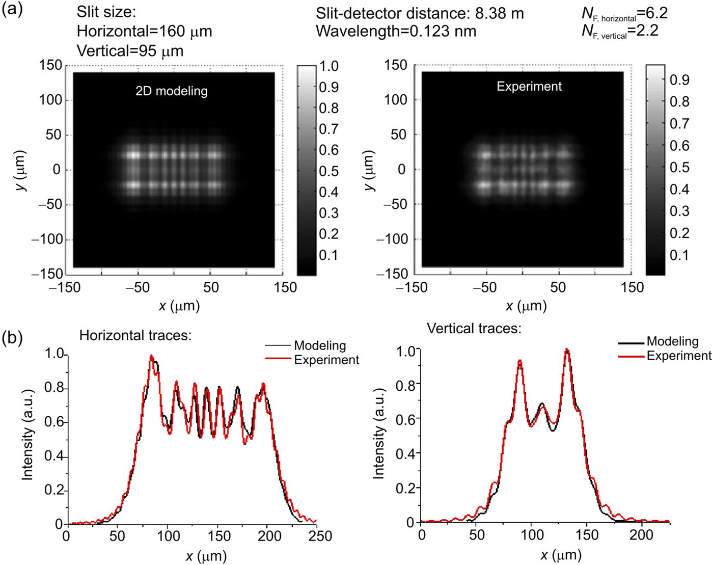
M. Ruiz-Lopez, A. Faenov, T. Pikuz, N. Ozaki, A. Mitrofanov, et al., Coherent X-ray beam metrology using 2D high-resolution Fresnel-diffraction analysis, J. Synchrotron Radiat. 24 (2017) 196–204.10.1107/s1600577516016568
|
Figures(14)


 DownLoad: